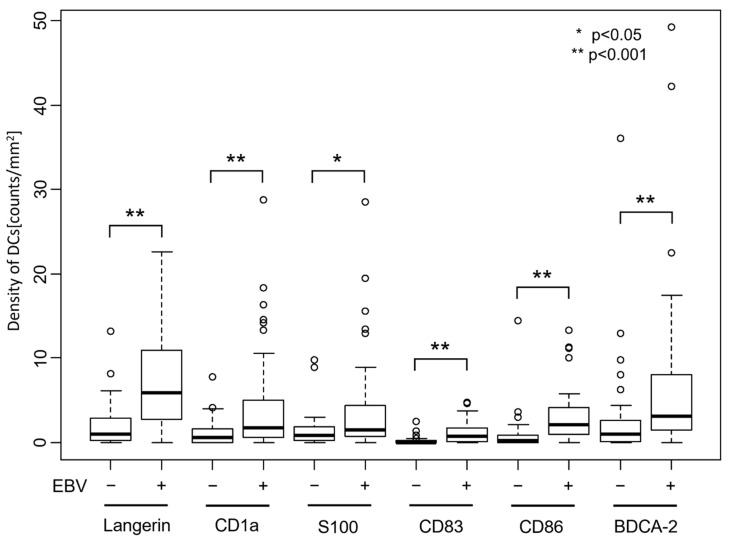
Figure 5.
Number of dendritic cells that infiltrated gastric cancer tissue. Dendritic cells expressing each DC marker were counted, and the number per tumor area is shown. The number of DCs is significantly higher in EBVaGC than in EBV-negative gastric cancer for any of the DC markers (p < 0.001 for Langerin, CD1a, CD83, CD86, and BDCA-2. p < 0.05 for S100, determined by using Wilcoxon rank sum test).

