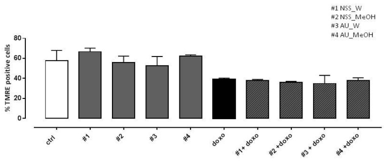
Figure 5.
Effect of NSS and AU extracts on doxo-induced mitochondrial membrane depolarization. NSS and AU extracts (50 µg/mL) were administered alone for 24 h or 4 h before doxo (1 µM). The mitochondrial membrane potential was evaluated by flow cytometry analysis with Tetramethylrhodamine ethyl ester (TMRE), a cell-permeant, positively-charged, red-orange dye which penetrates and accumulates in the mitochondria in inverse proportion to the membrane potential. The low value of percentage of TMRE+ cells means that the TMRE dye was not trapped in the mitochondrial membrane due to its depolarization. Data were expressed as mean ± SD of fluorescence intensity of at least three independent experiments (N = 6).

