Figure 1.
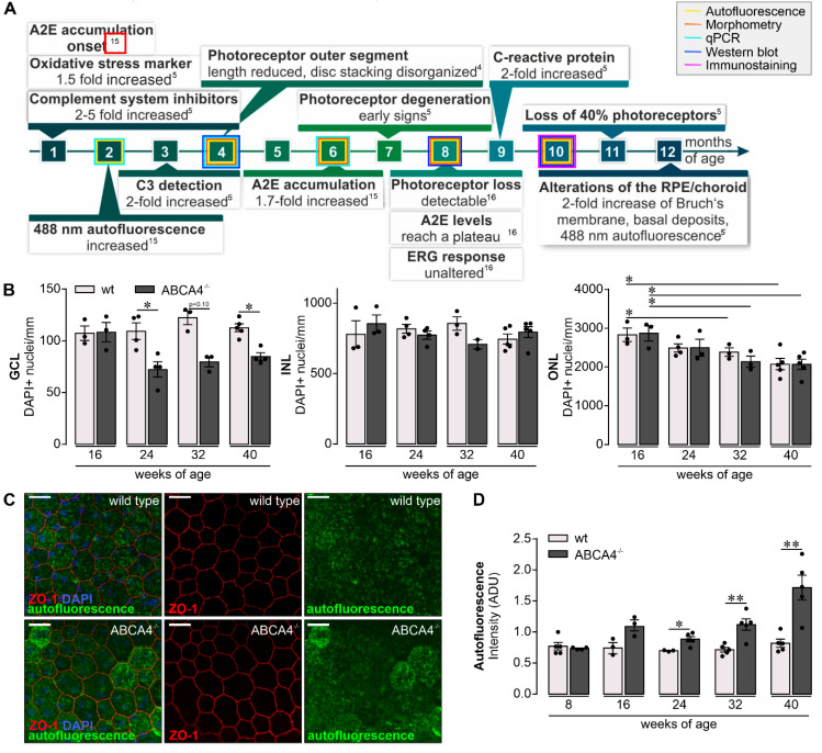
Retinal phenotype in the investigated albino ABCA4−/− strain. (A) Subsumed retinal phenotype of albino ABCA4−/− mice based on published studies [4,5,15,16]. ERG, electroretinogramm; ONL, outer nuclear layer; A2E, N-retinylidene-N-retinylethanolamine; C3, complement factor 3. Experiments performed in the course of the present study at different time points are highlighted by frames in respective colors. (B) A decrease in the number of DAPI+ cell nuclei in the ganglion cell layer (GCL) is observed in ABCA4−/− mice aged 24, 32 and 40 weeks but not in the albino wild-type retina (data were obtained previously) [19]. Cell numbers in the retinal inner nuclear layer (INL) were stable in aging ABCA4−/− mice. Photoreceptor density, as determined by quantification of DAPI+ cell nuclei in the outer nuclear layer (ONL), was significantly decreased in ABCA4−/− and wild-type mice at 32 weeks of age and older [19]. (C) The integrity and autofluorescence of the retinal pigment epithelium (RPE) were investigated in fixed, flat-mounted eyecup preparations after careful removal of overlaying retinae. ZO-1 labeling delineates tight junctions formed by intact RPE cells. Autofluorescence was detected upon excitation with a 488 nm laser and revealed enhanced signals at 40 weeks of age from the RPE of ABCA4−/− mice as compared to the RPE from wild-type littermates. Scale bars, 20 µm. (D) Mean gray values for autofluorescence were measured over the whole scan field. (B,D). Bars represent mean values ± SEM from 2 to 5 animals. * p < 0.05, ** p < 0.01, Mann–Whitney U-test (two-tailed). ADU, arbitrary digital units.

