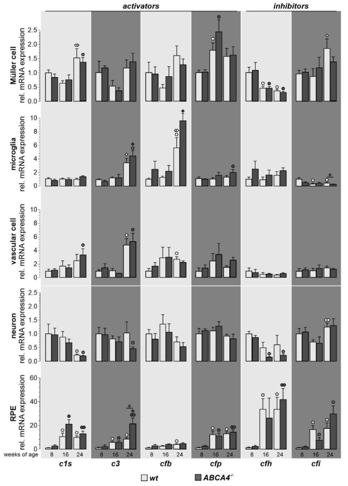
Figure 2.
Comparison of complement component expression between retinal cell types of wild-type albino and ABCA4−/− mice. Complement expression analysis of c1s, c3, cfb, cfp, cfh and cfi by qRT-PCR was performed on Müller cells, microglia, vascular cells, neurons and RPE from 8-, 16- and 24-week-old ABCA4−/− mice and compared to previously published wild-type data [19]. We found a significantly enhanced expression of c3 in RPE cells and decreased expression of cfi in microglia cells compared to wild-type controls. Most other age-dependent changes in complement expression were similar in both mouse strains [19]. Bars represent mean values ± SEM of cells purified from four to six animals. Mann–Whitney U-testing was performed on all data (*p < 0.05. White circle (◦): significant difference compared to the expression level at eight weeks of age in wt; black circle (●): significant difference compared to the expression level at eight weeks of age in ABCA4−/− mice; white diamond (◊): significant difference compared to the expression level at 16 weeks of age in wt animals; black diamond (♦): significant difference compared to the expression level at 16 weeks of age in wt animals. ◦/●/◊/♦: p < 0.05; ●●/◊◊: p < 0.01).

