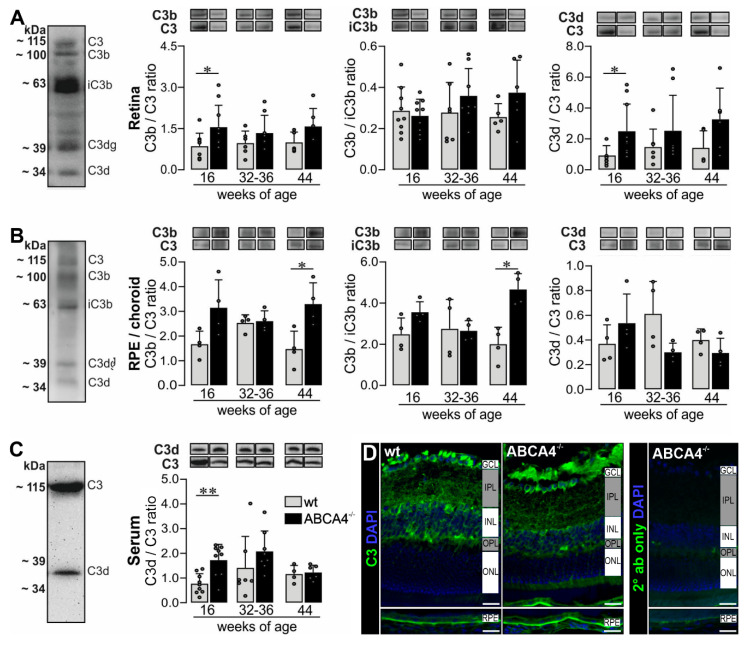
Figure 3.
Western blot analysis and immunostaining of complement factor C3 in the RPE/choroid, retina and serum of wild-type albino and ABCA4−/− mice. (A,B) Western blot analysis under reduced condition using an anti-C3 α-chain antibody showed bands for C3, C3b, iC3b, C3dg and C3d fragments in the (A) retina and (B) RPE/choroid. (A) At 16 weeks of age, we detected a significant increase of C3b to C3 and C3d to C3 ratio in the retina of ABCA4−/− mice compared to wild-type mice. (B) The ratio of C3b to C3 and C3b to iC3b was significantly increased in the RPE/choroid of ABCA4−/− mice compared to wild-type mice at 44 weeks of age. (C) C3 and C3d fragments were detected in the serum using the C3d-fragment-specific antibody. The C3d to C3 ratio was increased at 16 weeks of age in ABCA4−/− mice. (D) Immunofluorescence staining using an anti-C3 (green/gray) antibody showed an increase of C3 fragments in the RPE in albino ABCA4−/− mice compared to wild-type mice. Cell nuclei were counterstained with DAPI (blue). Stainings were performed on sections from at least three mice per genotype and representative images were chosen. As a negative control, sections were incubated with the secondary antibody (2° ab) only. GCL, ganglion cell layer; IPL, inner plexiform layer; INL, inner nuclear layer; OPL, outer plexiform layer, ONL, outer nuclear layer. Scale bars, 20 µm. (A–C) Bars represent mean values ± SEM from 4 to 10 animals. * p < 0.05, ** p < 0.01, Mann–Whitney U-test.

