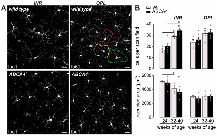
Figure 5.
Mild microglia activation in ABCA4−/− mice. (A,B) Microglia were quantified in the inner retinal layers (INR), such as the ganglion cell layer and inner plexiform layer, and additionally in the outer plexiform layer (OPL) on the basis of Iba1 labeling in mice of the indicated age in ABCA4−/− mice and compared to previously published data of wild-type albino mice [19]. (A) The area occupied by the widely branched processes of a single microglia was measured as exemplarily depicted by the dashed circles of different colors for the OPL microglia in a retina from a wild-type animal. Scale bars, 20 µm. (B) Bars represent mean values ± SEM from 2‒4 animals. * p < 0.05, Mann–Whitney U-test. ADU, arbitrary digital units.

