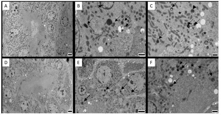
Figure 4.
Cortical renal cells from proximal convoluted tubules from (A) control broiler chicken; (B) broiler chicken in the experimental group exposed to 0.1 mg citrinin (CIT)/kg feed during 21 days; (C) broiler chicken in the experimental group exposed to 3 mg CIT/kg feed during 21 days; (D) control laying hen; (E) laying hen in the experimental group exposed to 0.1 mg CIT/kg feed during 21 days; (F) laying hen in the experimental group exposed to 3.5 mg CIT/kg feed during 21 days. Degenerated mitochondria are present in all treated animals (arrows); note remaining cristae inside the vesicles (arrowhead). The stars indicate examples of degenerating mitochondria, with increased space in between the cristae. Magnification: 2500×. Scale bars: (A,D,E):2 µm; (B,C,F): 1 µm.

