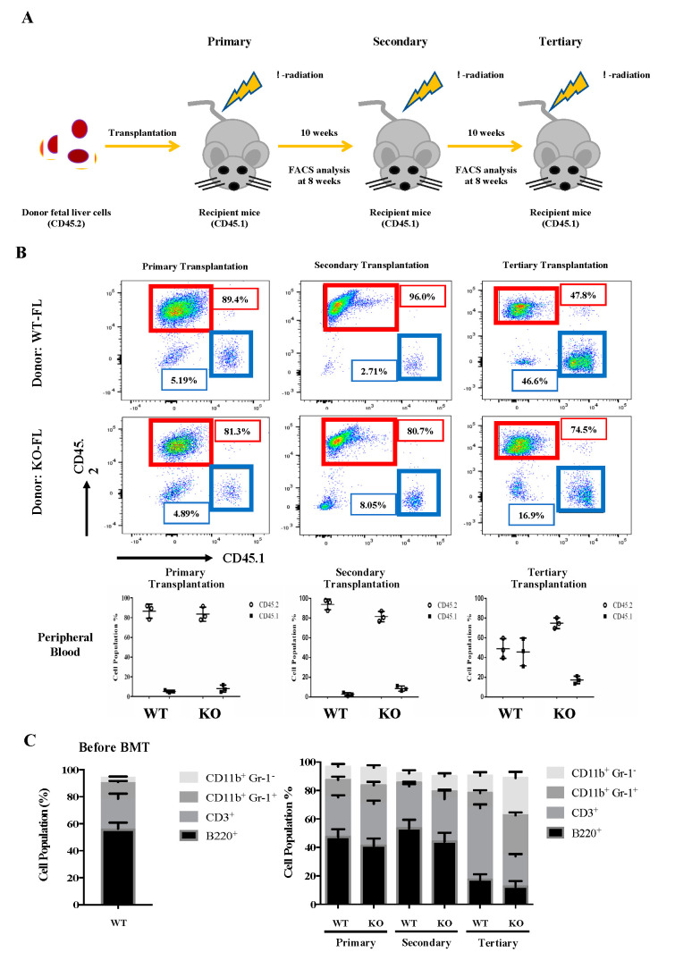
Figure 3.
Serial transplantation of fetal liver cells from WT and Eklf −/− E14.5 embryos. (A) Strategy of the experiments. The fetal liver cells (CD45.2) were transplanted into lethally irradiated recipient mice. The percentages of donor/recipient chimerism of the peripheral blood were analyzed by flow cytometry at 8 weeks after each transplantation. (B) Flow cytometric analysis of the donor/recipient chimerism of the peripheral blood of the recipient mice. Representative FACS plots are shown in the upper six panels. Statistical analysis is shown in the lower three histograms. The different cell populations were normalized to the total number of CD45.1 and CD45.2 cells. The data represent mean ± S.D (primary transplantation, n = 3; secondary transplantation, n = 4; tertiary transplantation, n = 8) (C) Percentages of donor-derived lineage repopulations of T cells (CD3+), B cells (B220+), monocytes (CD11B+ Gr1−) and granulocytes (B11b+ Gr-1+) in the peripheral blood of recipient mice. The data represent mean ± S.D (primary transplantation, n = 3; secondary transplantation, n = 4; tertiary transplantation, n = 8).

