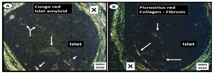
Figure 13.
Islet amyloid and islet fibrosis in human islets co-occur. (Panel (A)) depicts Congo red staining (stains amyloid) viewed with crossed polarized light and birefringent appearance at the peri-islet (X) and intra-islet regions (arrows). (Panel (B)) illustrates islet fibrosis (collagen I and III) staining with picrosirius red and viewed with crossed polarized light of pancreatic islet at the peri-islet (X) and intra-islet regions (arrows). These images demonstrate that islet amyloid and islet fibrosis co-occur within human pancreatic islets when stained specifically for amyloid (Congo red) and fibrosis (picrosirius red) in a female patient who died of an acute myocardial infarction [54]. Specific staining procedures by pathologists often reveal what standard hematoxylin and eosin (H&E) staining do not in regard to conditions such as islet amyloid and fibrosis. Magnification ×100 objective with oil immersion in Panels (A,B).

