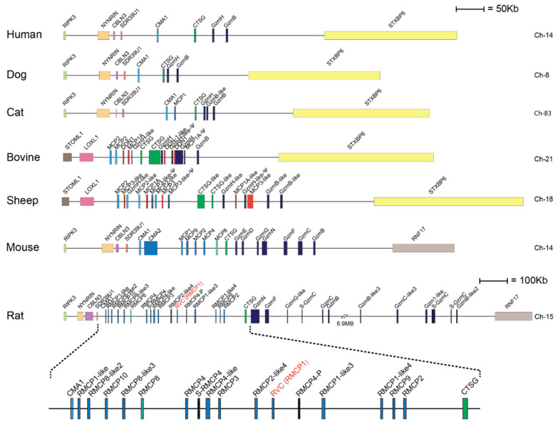
Figure 1.
The mast cell chymase locus with their bordering genes of seven different mammals. The species is given to the left side of the figure and the chromosome on which the chymase locus is located to the right. The genes for the serine proteases are shown at double height for more easy identification with their names as found in the database. Granzymes are color coded in dark blue, α-chymases in light blue and β-chymases as blue with a darker tint. MCP-8-related genes are in cyan, duodenases in red and Cathepsin G in green. The location of rat vascular chymase is indicated with red text.

