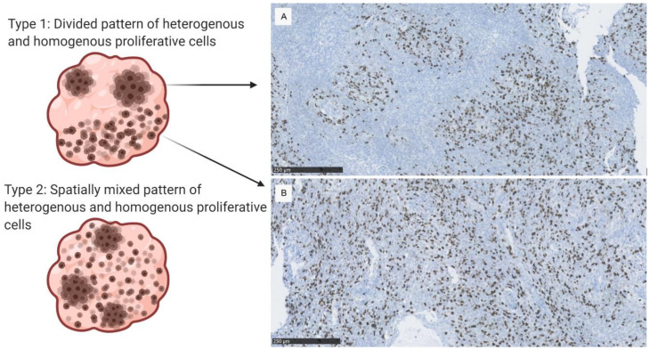
Figure 2.
Anaplastic meningioma from the cohort, Ki-67 stain. (A) Hot spot pattern with distinct areas of Ki-67 positive cells. (B) A homogenous proliferative pattern. The A and B patterns can be mixed within the tumor resulting in either a type 1 or type 2 proliferative pattern (shown on the left), which were observed in the 24 malignant meningioma samples.

