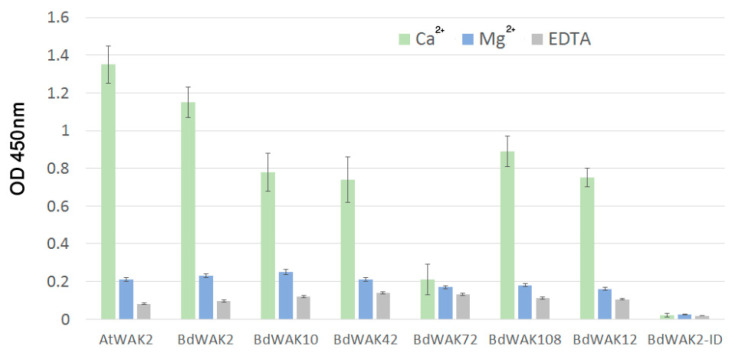
Figure 4.
ELISA assay for interaction of recombinant WAK extracellular domains with PGA. PGA prepared in 0.5 mM Ca2+/150 mM Na+ Tris buffer (green), in 0.5 mM Mg2+/150 mM Na+ Tris buffer in which Ca2+ is replaced by Mg2+ (blue) or in 0.5 mM Ca2+/150 mM Na+ Tris buffer + 5 mM EDTA (grey) was used to coat ELISA plates. BdWAK2-ID is the intracellular domain of BdWAK2 included as a negative control. The presence of WAK extracellular domain remaining bound to PGA was detected by anti-6× His antibody binding (and subsequent application of secondary mouse-HRP antibody) to the His-tagged extracellular domain, and quantified by the colour change of TMB (a HRP substrate) at OD 450 nm. Each measurement was performed in triplicate. Error bars indicate standard error.

