Figure 1.
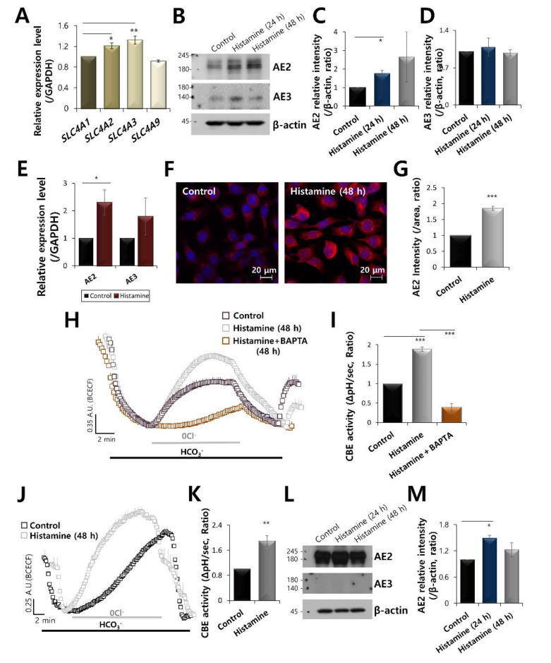
AE2 is activated by the stimulation of histamine in keratinocytes. (A) mRNA expression of SLC4 family receptors (SLC4A1, A2, A3, and A9) in HaCaT keratinocytes. (B) The protein expression of AE2, AE3, and β-actin during histamine treatment at 24 and 48 h in HaCaT keratinocytes. The β-actin was used as a loading control. Analysis of AE2 expression (C) and AE3 expression (D) with or without histamine in HaCaT cells. The bars indicate the mean ± SEM of data (* p < 0.05, n = 4). (E) mRNA expression of AE2 and AE3 with or without histamine stimulation in HaCaT cells (* p < 0.05). (F) Immunostaining of AE2 (red) and nucleus (DAPI, blue) in the presence of 500 nM histamine at 48 h. (G) The bars indicate the mean ± SEM of the AE2 membrane intensity determined from three experimental replicates (*** p < 0.001, n = 3). (H) CBE activity of HaCaT keratinocytes with (grey open square) and without (control, black open square) 500 nM histamine and with co-stimulation of 500 nM histamine and 10 μM BAPTA-AM (orange open square) at 48 h. Averaged traces were represented. (I) Analysis of CBE activity. The bars indicate the means ± SEM of the number of experiments (*** p < 0.001, n = 4). (J) CBE activity of primary keratinocytes with (grey open square) and without (control, black open square) 500 nM histamine at 48 h. (K) Analysis of CBE activity of primary epidermal keratinocytes. The bars indicate the means ± SEM of the number of experiments (** p < 0.01, n = 3). (L) The protein expression of AE2, AE3, and β-actin during histamine treatment at 24 and 48 h in primary epidermal keratinocytes. The β-actin was used as a loading control. (M) Analysis of AE2 expression with or without histamine stimulation in primary epidermal keratinocytes. The bars indicate the mean ± SEM of data (* p < 0.05, n = 4).

