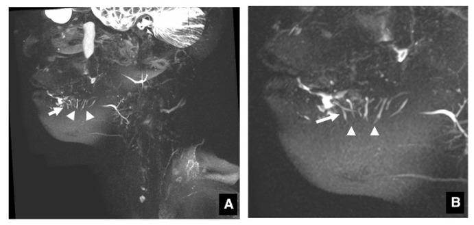
Figure 1.
MR sialography of sublingual gland ducts. Extraglandular portions of the typical sublingual gland ducts in MR sialography ((A): overall image, (B): enlarged image of the sublingual gland area) can be identified as many bright, homogeneous, ascending linear structures (arrows) in continuity with the sublingual glands (arrowheads).

