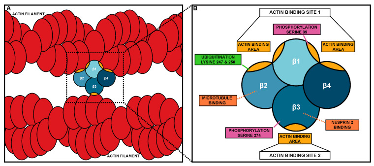
Figure 1.
Fascin bundling mechanism and actin-binding domains. (A) Schematic of Fascin bundling two actin filaments (red). (B) Schematic of the domains and binding site of Fascin. The four β-trefoil domains of Fascin are in different shades of blue. Actin-binding areas of Fascin are in gold. In B, the different protein interaction sites and post-translational modifications are labeled.

