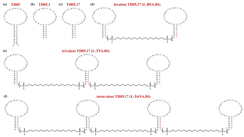
Figure 7.
Schematic representation of the secondary structure of the monomeric and dimeric mIgM-targeting aptamers: TD05 (a), TD05.1 (b), TD05.17 (c), bivalent TD05.17, i.e., L-BVA.8S (d), trivalent TD05.17, i.e., L-TVA.8S (e) and tetravalent TD05.17, i.e., L-TetVA.8S (f). Nucleobases in red color are LNA residues. Figures were redrawn from Mallikaratchy et al. [185].

