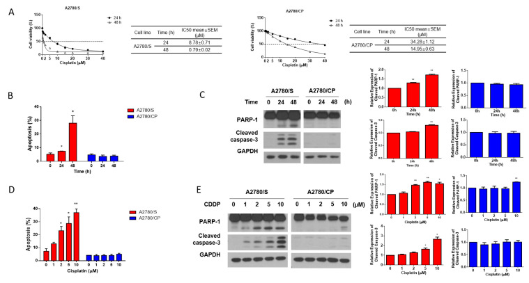
Figure 1.
(A) A2780/S and A2780/CP cells were treated with the indicated concentrations of cisplatin for 24 and 48 h. Cell viability was evaluated by MTT assay and the best-fit curves were generated to determine IC50 values of A2780/S and A2780/CP cells; (B) A2780/S and A2780/CP cells were treated with cisplatin (2 μM) for 24 and 48 h. Cell apoptosis was detected by flow cytometry using Annexin V staining; (C) The expression of apoptosis-related proteins was determined by Western blotting; (D) A2780/S and A2780/CP cells were treated with cisplatin in different concentrations (0 to 10 μM) for 48 h. Cell apoptosis was detected by flow cytometry using Annexin V staining; (E) The expression of apoptosis-related proteins was determined by Western blotting. Results are shown as mean ± SEM (* p ≤ 0.05, ** p ≤ 0.01).

