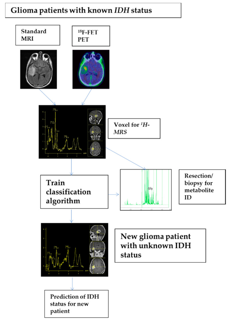
Figure 4.
Detailed process description of analysis pipeline as it could be applied in clinical praxis. First, standard MRI and 18F-FET PET were applied for voxel planning. Second, in selected voxels 1D 1H MRS spectra of patients with known IDH status were acquired. Next, a classification algorithm, namely a linear support vector machine, was trained on all spectra of the training set implementing a nested leave-one-out cross-validation approach. Finally, once training is completed new spectra of patients with unknown IDH status may be presented to the algorithm for prediction of IDH status.

