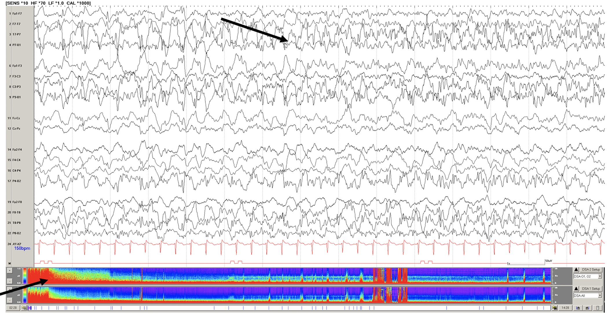
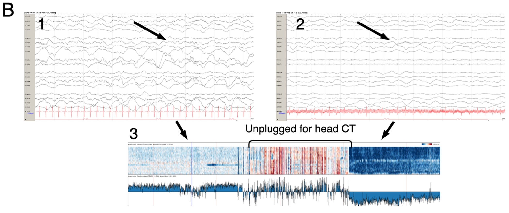
Figure 1: Examples of EEG tracings for patients supported on ECMO.


A. Raw EEG tracing of a 7 year old with respiratory failure requiring VA ECMO support. Continuous video EEG monitoring initiated soon after cannulation revealed status epilepticus (black arrows on raw EEG tracing and density spectral array) and subsequent seizures during recording. B. Raw EEG (1 and 2) and asymmetry index from quantitative EEG (3) of a 1 year-old child on VA ECMO support after near-drowning. While unplugged from EEG recording for head CT the patient suffered a right hemispheric stroke. (1) Raw EEG tracing with symmetric sleep spindles corresponding to normal asymmetry index. Upon return from CT suite, the patient demonstrated asymmetry of sleep spindles (2) and dramatically increased relative power or activity (dark blue in 3) on the left and not right hemisphere.
