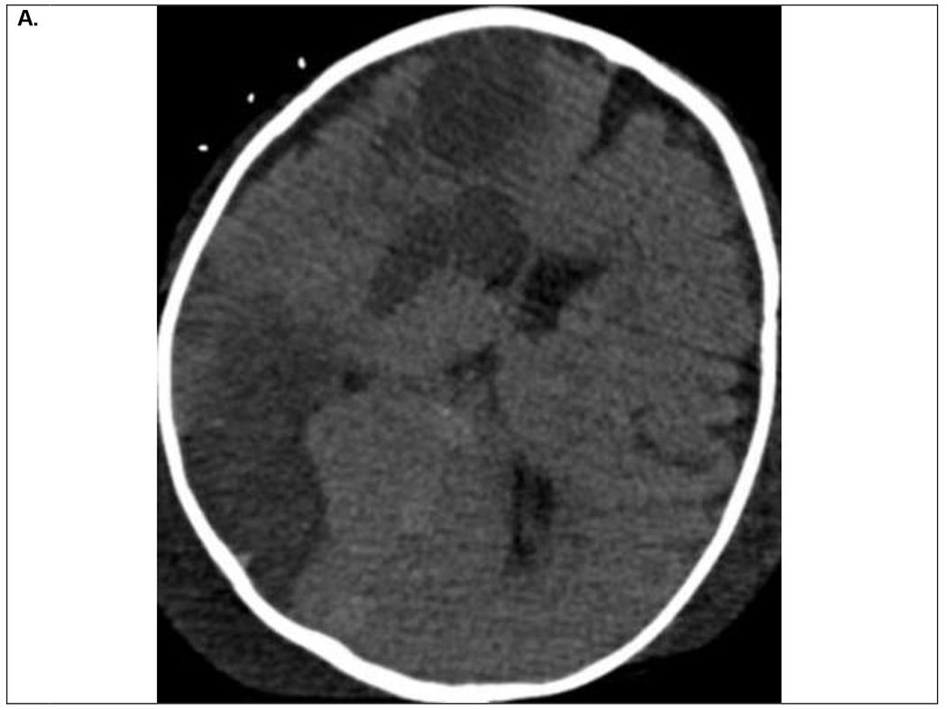
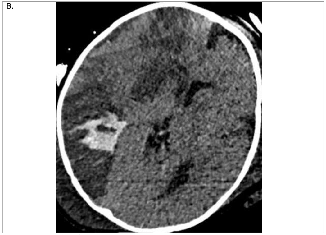
Figure 2.


9 month old infant on VA ECMO support for refractory septic shock and multiorgan dysfunction. Within first 24 hours of ECMO course, the patient developed clinical signs of right sided facial venous congestion and asymmetric slowing on EEG, initially thought to be secondary to superior vena cava obstruction by the ECMO venous drainage cannula. A. Head CT on ECMO day 3 demonstrated multifocal ischemic infarcts within the right hemisphere for which his systemic anticoagulation was temporarily held. B. Follow up head CT on ECMO day 5 demonstrated evolution of the ischemic infarcts and hemorrhagic conversion within the posterior ischemic infarct. He was decannulated on ECMO day 9 and discharged home on hospital day 68 with residual left hemiparesis.
