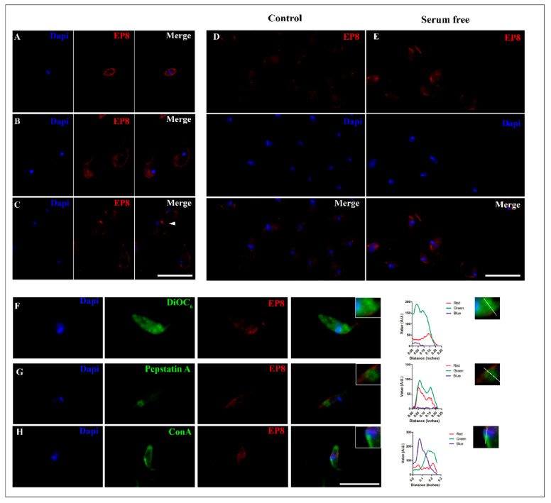
Figure 5.
Immunofluorescent subcellular localization of the T. cruzi PS-like protein. A single z-plane image of a representative parasite of different forms of T. cruzi, (A) amastigote (B) trypomastigote and (C) epimastigote immunolabeled with anti-EP8 (red) and DAPI to label nuclei and kinetoplast (blue). White arrowhead shows an immunolabeled concentration signal near the flagellar pocket. Increased signal of PS-like protein in epimastigotes from control conditions in 10% FBS (Control; (D)) to serum deprivation for 24 h (E). Cytolocalization of EP8 with the ER stained with DiOC6 (F), intracellular vesicles marked with Cathepsin B (G) and the flagellar pocket (H). Fluorescence intensity plot of each channel (Red, Green, and Blue) were performed, tracing a line from the posterior to the anterior region of the parasite for each organelle dye. Scale bar = 10 µm.

