Figure 1.
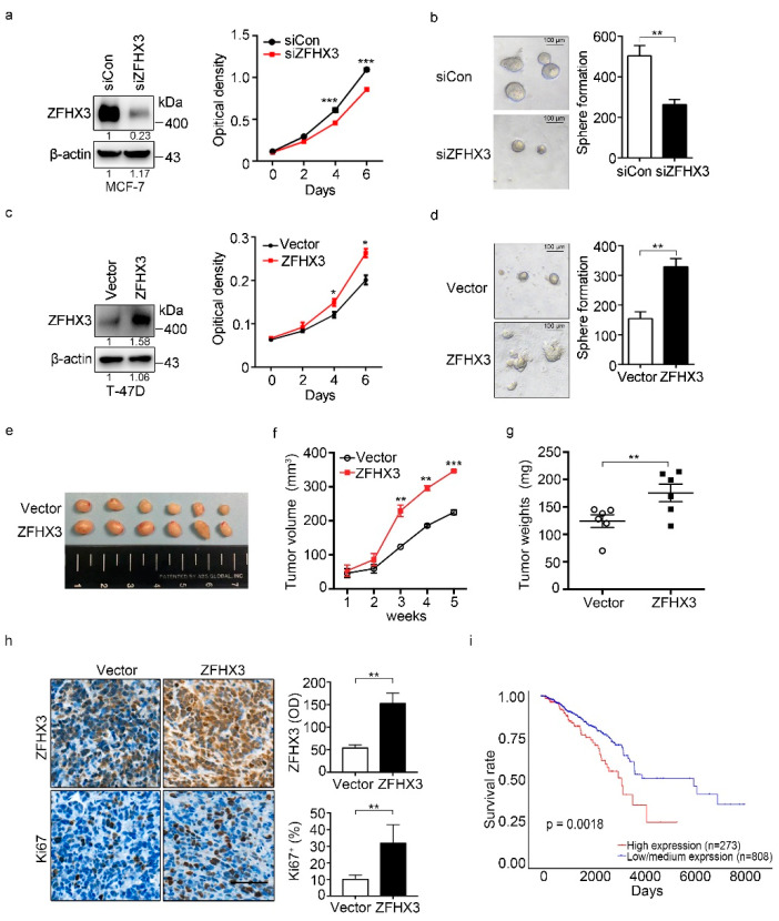
ZFHX3 enhances colony formation and tumorigenicity of ER+ breast cancer cells. (a,b) ZFHX3 silencing by RNAi inhibited colony formation in 2D culture (a), as determined by the sulforhodamine B (SRB) assay, and sphere formation in Matrigel (b), as indicated by representative images of spheres (left) and the numbers of spheres with a diameter >75 µm (right) in MCF-7 cells. siCon, control siRNA; siZFHX3, siRNA against ZFHX3. (c,d) Ectopic expression of ZFHX3 promoted colony formation in the 2D SRB assay (c) and 3D sphere formation in Matrigel (d) in T-47D cells. (e–g) ZFHX3 expression in T-47D cells also promoted their subcutaneous tumor formation in nude mice, as indicated by tumor images (e), tumor growth curve (f), and tumor weight (g). (h) Enhanced tumor growth by ZFHX3 was accompanied by increased cell proliferation, as indicated by representative images of IHC staining of ZFHX3, Ki67 in xenograft tumors, and the quantification of ZFHX3-positive cells (up), Ki67-positive cells (down). Scale bar, 50 µm. (i) Higher mRNA levels of ZFHX3 are associated with worse survival in patients with breast cancer in the TCGA database, as analyzed by survival analysis. * p < 0.05; ** p < 0.01; *** p < 0.001. Ratios of protein band intensities to those of their loading controls, with the control sample’s normalized to 1, are shown under bands in western blots (a,c). Uncropped western blot images are available in Supplementary Figure S7.

