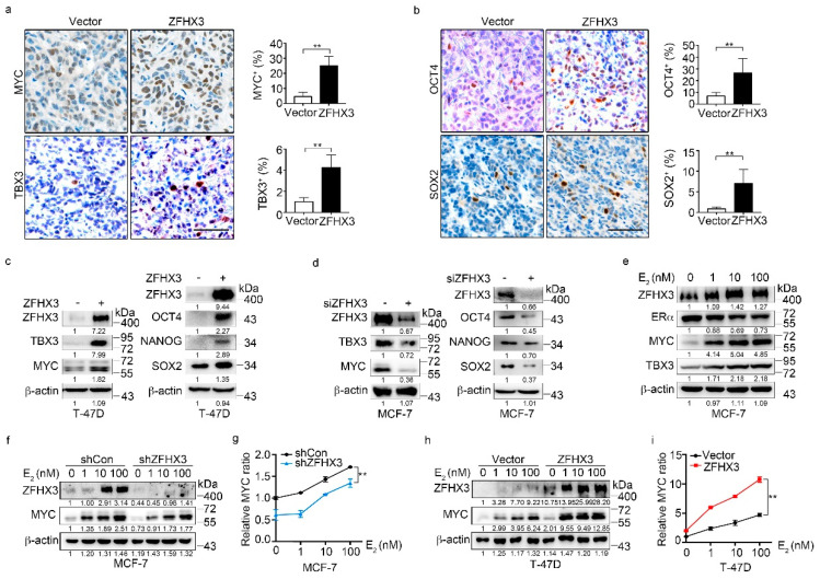
Figure 3.
ZFHX3 induces the expression of MYC and TBX3 in ER+ MCF-7 and T-47D breast cancer cells. (a,b) Detection of MYC, TBX3, OCT4, and SOX2 by IHC staining in xenograft tumors of T-47D cells ectopically expressing ZFHX3, as shown by representative IHC images (left) and quantification of cells with positive staining (right). Scale bar, 50 µm. (c,d) Ectopic expression of ZFHX3 in T-47D cells increased (c), while ZFHX3 silencing in MCF-7 cells decreased (d), the expression of MYC, TBX3, OCT4, NANOG, and SOX2, as detected by western blotting analysis. (e) Estrogen (E2) increased MYC and TBX3 in MCF-7 cells, as detected by western blotting. (f–i) ZFHX3 silencing in MCF-7 cells attenuated, while ectopic expression of ZFHX3 in T-47D cells enhanced, E2-mediated MYC expression, as detected by western blotting (f,h) and quantified by relative MYC ratio (g,i). Cells were treated with E2 for 24 h at the indicated concentrations. siCon, control siRNA; siZFHX3, siRNA against ZFHX3. ** p < 0.01. (a,b) IHC staining was done in xenograft tumors. (c–i) cells were cultured in 2D plastic surface and examined with western blotting. Ratios of protein band intensities to those of their loading controls, with the control sample’s normalized to 1, are shown under western blot bands (c–f,h). Uncropped western blot images are available in Supplementary Figure S8.

