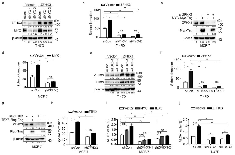
Figure 5.
MYC and TBX3 play a role in ZFHX3-mediated sphere formation in Matrigel and ALDH+ cell population in ER+ MCF-7 and T-47D breast cancer cells. (a,b) In T-47D cells ectopically expressing ZFHX3, MYC silencing, as confirmed by western blotting in 2D culture (a), and culture in Matrigel (3D culture) that attenuated sphere formation in both ZFHX3 and vector control groups (b). (c,d) In MCF-7 cells with ZFHX3 silencing, MYC ectopic expression, as confirmed by western blotting in 2D culture (c), and culture in Matrigel (3D culture) that did not significantly increase sphere number (d). (e,f) In T-47D cells ectopically expressing ZFHX3, TBX3 silencing, as confirmed by western blotting in 2D culture (e), and culture in Matrigel (3D culture) that attenuated sphere formation (f). (g,h) In MCF-7 cells with ZFHX3 silencing, TBX3 ectopic expression, as confirmed by western blotting in 2D culture (g), and culture in Matrigel (3D culture) that did not significantly increase sphere number (h). (i,j) ALDH+ cells were then detected by flow cytometry in MCF-7 cells with ZFHX3 silencing and MYC or TBX3 overexpression (i) and T-47D cells with ZFHX3 overexpression and MYC or TBX3 knockdown (j). ns, not significant; * p < 0.05; ** p < 0.01. Cells were cultured on a 2D plastic surface. siCon, control siRNA; siZFHX3, siRNA against ZFHX3. Ratios of protein band intensities to those of their loading controls, with the control sample’s normalized to 1, are shown under western blot bands (a,c,e,g). Uncropped western blot images are available in Supplementary Figure S9.

