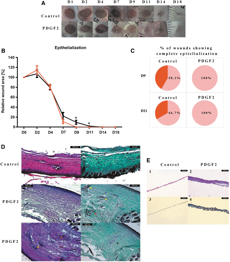
Figure 6.
Representative images of dorsal skin wounds in mice (A), wound epithelialization (B), and the percentage of wounds showing complete epithelialization (C) in mice treated with PDGF2. Histological analysis of wound healing: (D) images of skin samples at day 21 after injury and stained with hematoxylin and eosin (on the left) or Masson's trichrome (on the right). Yellow arrows indicate hair follicles, and red arrows indicate glands. Black arrows show areas of cells with a distinct morphology. (E). Images of epithelial membrane samples at day 4 postinjury stained with hematoxylin and eosin (1–2) or Masson's trichrome (3–4).

