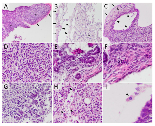
Figure 1.
Experimental mouse teratoma morphology. (A) teratoma consisting mostly of undifferentiated tissue (asterisk—cords and sheets of mainly monomorphic cells with high mitotic activity and without any visible forming structure), while on the surface clearly differentiated tissue is present (arrow—morphological cylindrical epithelial cells with eosinophilic cytoplasm and pseudostratified epithelial cells) (HE, 200×); (B) teratoma composed of differentiated tissue (arrow—surface epithelial cells, arrowhead—cystic structure lined by thin flattened epithelia), while the inside is mostly made up of UTC (asterisk) (HE, 200×); (C) the teratoma composed of all three germ cell-derived lines, mesodermal (asterisk), endodermal (arrowhead) and ectodermal (arrow) (HE, 200×); (D) high magnification of UTCs (HE, 400×); (E) high magnification of CSCLC (asterisk) (HE, 400×); (F) high magnification view of hematopoiesis (asterisk) (HE, 400×); (G) high magnification of lymphocytes (asterisk) (HE, 400×); (H) high magnification of cells in the process of apoptosis (arrowhead) (HE, 400×); (I) high magnification of a cell in the process of mitosis (arrowhead) (HE, 600×).

