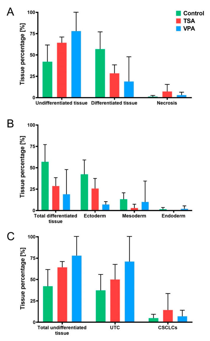
Figure 5.
Teratoma tissue distribution. (A) Teratoma tissue components across treatment groups, divided into undifferentiated tissue, differentiated tissue and necrotic tissue. (B) Tissue differentiation according to germ layer of origin, with percentage of differentiated tissue depicted as belonging to ectodermal, mesodermal or endodermal tissue across treated groups. (C) Stratification of undifferentiated teratoma components into undifferentiated teratoma cells (UTC) and cancer stem cell-like cells (CSCLCs). Values represented are means with 95% CI.

