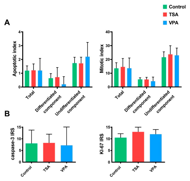
Figure 6.
Analysis of apoptosis and proliferation. (A) Apoptotic and mitotic indices have been measured in in vitro-cultured teratoma HE slides as total number of apoptotic or mitotic cells in differentiated and undifferentiated teratoma tissue. Values represented are means with 95% CI. (B) IHC analysis of apoptosis and proliferation in in vitro-treated teratoma. Proliferation has been analyzed based on Ki-67 protein expression, while apoptosis by caspase-3 expression. Values represented are means with 95% CI.

