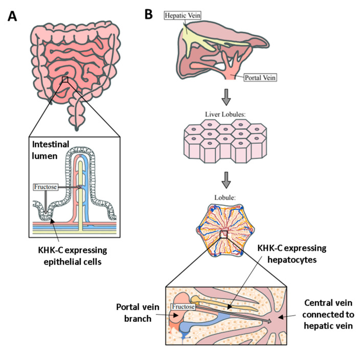
Figure 3.
Anatomical difference between the liver and intestine for fructose catabolism. (A) In the intestine, there is a single-layer of KHK-C expressing epithelial cells between the intestinal lumen and blood vessel (fructose diffusion is perpendicular to the cell layer). This structure limits the fructose catabolic capacity when the delivered fructose dose is high. (B) In contrast, in the liver, there are numerous KHK-C-expressing hepatocytes lining the portal-to-hepatic circulation. Hepatocytes are also metabolically highly active to efficiently assimilate fructose carbons via gluconeogenesis and fat synthesis.

