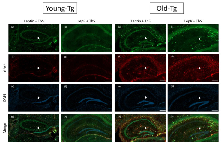
Figure 3.
Leptin and LepR with Thioflavin-S and glial fibrillary acidic protein (GFAP) staining on the hippocampi of young (10–12 week) and old (48–52 week) 5XFAD (Tg) mice. Amyloid plaques stained by thioflavin-S (ThS) are indicated by white arrows. Plaques were more prevalent in the hippocampi of old-Tg (i,j,o,p) compared to young-Tg mice (a,b,g,h). Leptin and LepR was detected in the hippocampus of young-Tg (a and b, respectively) and to a lesser extent in old-Tg mice (i and j, respectively). Both young- (c,d) and old-Tg (k,l) mice also had astrogliosis identified by GFAP staining. Furthermore, astrocytic expression of leptin (o) and LepR (p) with GFAP was observed in the hippocampi of old-Tg mice compared to young-Tg mice (g and h, respectively). Young- (e,f) and old-Tg (m,n) sections were counterstained with 4′,6-diamidino-2-phenylindole (DAPI). Scale bar = 200 μm.

