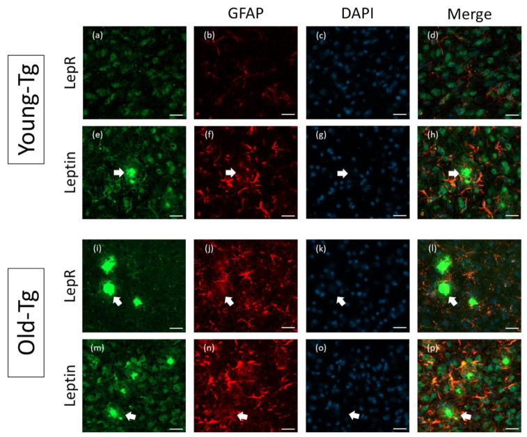
Figure 4.
Astrocytic expression of leptin and LepR in the cerebral cortex of young (10–12 week) and old- (48–52 week) 5XFAD (Tg) mice. Thioflavin-S-stained amyloid plaques (arrows) are present in the cerebral cortex of old-Tg and to a lesser extent in young-Tg mice. LepR and leptin was detected in neurons and astrocytes in the cerebral cortex of young- (a and e, respectively) and old-Tg (i and m, respectively) mice. Panels (b,f,j,n) depict astrocytes labelled with GFAP. Panels (c,g,k,o) depict nuclei staining with DAPI. Colocalization of LepR with GFAP (l) and leptin with GFAP (p) labelled astrocytes of old-Tg mice was observed. Co-expression of LepR (d) and leptin (h) with GFAP was also observed to a lesser extent in young-Tg mice. Scale bar = 20 μm.

