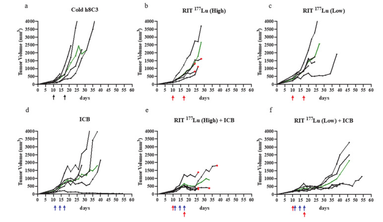
Figure 2.
Combination study of anti-PD1 mAb vs. RIT with 177Lu-h8C3 mAb. The mice in groups of five were treated with: (a) two doses of unlabeled (cold) h8C3 anti-melanin mAb on day 10 and 17 after S91 Cloudman tumor cell inoculation; (b) two doses of 200 μCi (High) of 177Lu-h8C3 mAb on day 10 and 17 (RIT 177Lu High); (c) two doses of 100 μCi (Low) of 177Lu-h8C3 mAb on day 10 and 17 (RIT 177Lu Low); (d) three doses of 250 μg anti-PD1 mAb on day 11, 14, and 17 (ICB); (e) three doses of 250 μg anti-PD1 mAb on day 11, 14, and 17 with two doses of 200 μCi (High) of 177Lu-h8C3 mAb on day 10 and 17 (RIT 177Lu High + ICB); (f) three doses of 250 μg anti-PD1 mAb on day 11, 14, and 17 with two doses of 100 μCi (Low) of 177Lu-h8C3 mAb on day 10 and 17 (RIT 177Lu Low + ICB). Day 0 is the day of tumor cell inoculation. Each black tumor volume curve represents a single animal; green line—median tumor volume in the group. Black, blue, and red arrows indicate the injection of cold antibody, anti-PD1 antibody, and RIT, respectively. Red dots indicate the death of the animal from radiotoxicity.

