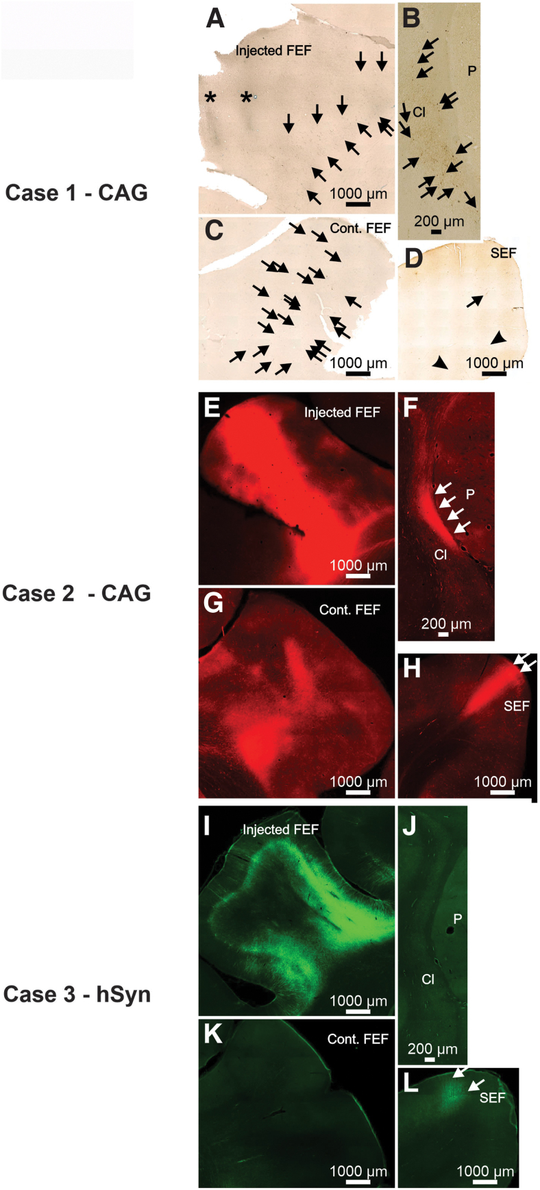
Figure 4.
Photomicrographs from Case 1 (A–D), 2 (E–H), and 3 (I–L) illustrating neuronal labeling provided by AAV2-retro-CAG (primate Cases 1 and 2) or AAV2-retrohSyn following injections into the FEF. Asterisk indicates the location of needle tracts. Arrows indicate locations of individual neurons in photomicrographs where it may not be obvious to the observer.

