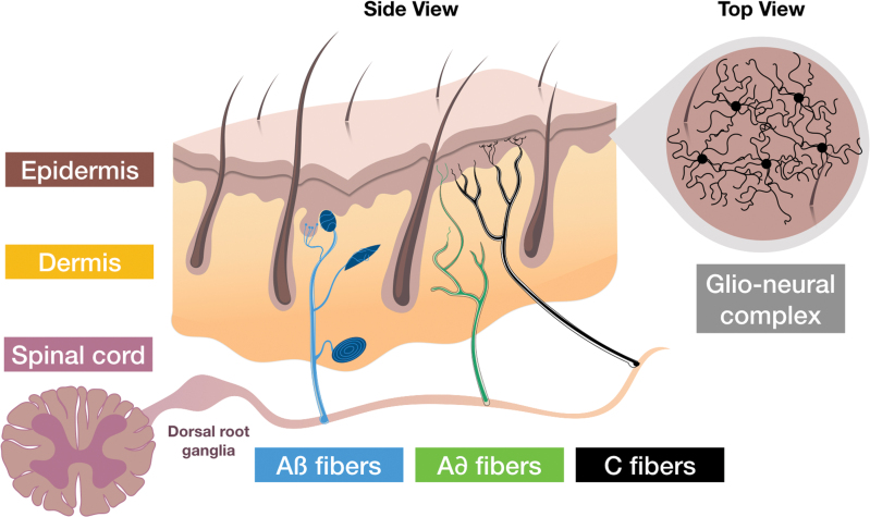
Figure 2.
Skin innervation. The sensory endings, which extend throughout all layers of the skin, transfer signals from mechanoreceptors, nociceptors, and thermoreceptors to the cell bodies located in the dorsal root ganglia. From there, stimuli, including itching, pain, and burning, are forwarded to specific areas in the brain through the spinal cord. Cutaneous sensory fibers are classified, according to diameter and speed of conduction, as Aβ, Aδ, and C fibers. Aβ fibers are fast and have large diameter, whereas C fibers are slow and have small diameter. Aβ and Aδ are myelinated by accompanying Schwann cells. The fragile C fibers are protected by nonmyelinating Schwann cells. Mechanical stimuli are detected by mechanoreceptors associated with sensory corpuscles (Meissner, Pacini, Ruffini corpuscles, and Merkel discs) through Aβ fibers, while pain and temperature are detected, respectively, by nociceptors and thermoreceptors through free endings of Aδ and C fibers. Filament-like protrusions associating nerve endings (C fibers) and mechanosensitive Schwann cell processes that extend into the epidermis, form the glio-neural complex (recently described in mice), a mesh-like structure that also participates in mechanical nociception.11 Color images are available online.

