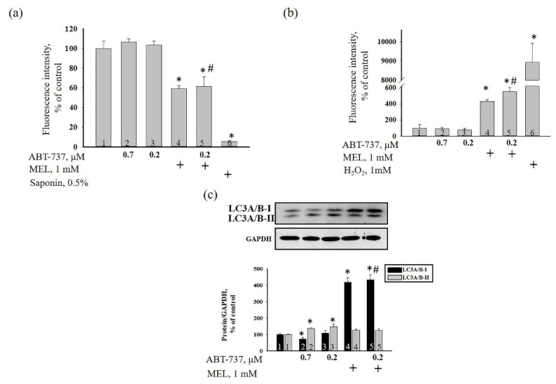
Figure 4.
The effect of MEL and ABT-737 on the membrane potential (ΔΨm) (a), ROS production (b) and in the level of autophagy marker LC3A/B (c) in HL-60 cells. Cells were treated with 0.7 µM ABT-737 (column 2), and 0.2 µM ABT-737 (columns 3) and 1 mM MEL (column 4), and MEL in combination with 0.2 µM ABT-737 (column 5); untreated cells (control, column 1). (a) the alteration of ΔΨm in our experimental conditions. Saponin (0.5%) was used as a positive control; (b) the alteration of ROS production in our experimental conditions. H2O2 (1 mM) was used as a positive control; (c) western blot of autophagy marker LC3A/B. The ratio of protein levels to GAPDH was used as a loading control. The protein level in the cell lysate without any additives served as a control (100%). “+” means the presence of MEL, Saponin and H2O2, respectively. The data are presented as the means ± S.D. of five separate experiments. * p < 0.05 significant difference in values compared with the corresponding control (untreated cells), # p < 0.05 significant difference in values relative to ABT-737 alone (0.2 µM, column 3).

