Figure 5.
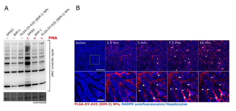
Protein kinase C inhibition by hepatocyte-directed PLGA-DY-635 (BIM-I) NPs. (A) DY-635 HepG2 pretreated with DMSO, free or encapsulated or BIM-I (200 nmol L−1) for 30 min and stimulated with PMA for 15 min. pPKC substrates were detected by Western blotting from total protein lysates and loading was evaluated by Coomassie staining of the gel. (B) Representative images from intravital microscopy of PLGA-DY-635 (BIM-I) NPs in the liver. Hepatocytes are detected through their strong NADPH auto-fluorescence and liver sinusoids can be identified by their negative staining (black). Lower panel depicts a zoom area (green square) of the upper panel. Some DY-635 accumulation in Kupffer cells (asterisks) was observed. DY-635-stained bile canaliculi (arrowheads) indicated the uptake and degradation of the NPs as well as elimination of DY-635. Scale bar: 0.1 mm (upper panel) and 0.025 mm (lower panel).

