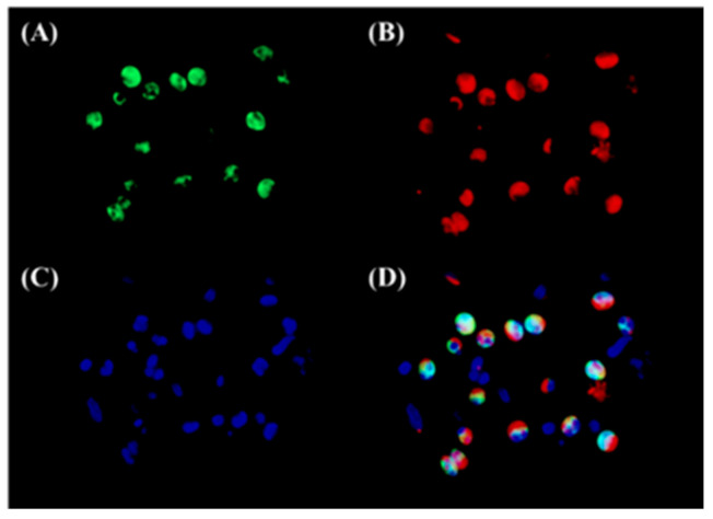
Figure 3.
Immunofluorescence of 15-LOX-1 and ECP in the sub-epithelial lesion of NP tissues from ECRS patients. An immunofluorescence assay was performed with anti-15-LOX-1 antibody (green fluorescence) (A), and anti-ECP mAb (red fluorescence) for eosinophils (B). Nuclei were counterstained with 4′,6-diamidino-2-phenylindole (DAPI); blue fluorescence, (C); and the signals were merged (D). The results are representative images from six different patients. Magnification: ×400.

