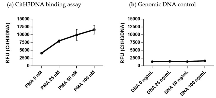
Figure 3.
Quantitative citrullinated histone H3 bound to DNA (CitH3DNA) binding assay for detection of NETs. High binding 96-well plates were coated with anti-histone H3cit antibodies. (a) Neutrophils were isolated from BM of myeloma-bearing mice and stimulated in vitro with indicated concentrations of PMA for 4 h. NET fractions were collected from each condition and 100 μL were loaded per well. (b) Indicated amount of genomic DNA was added per well. (a,b) Fluorescence of SYTOX Green nucleic acid stain was measured using a Victor X3 plate reader. RFU—relative fluorescence units.

