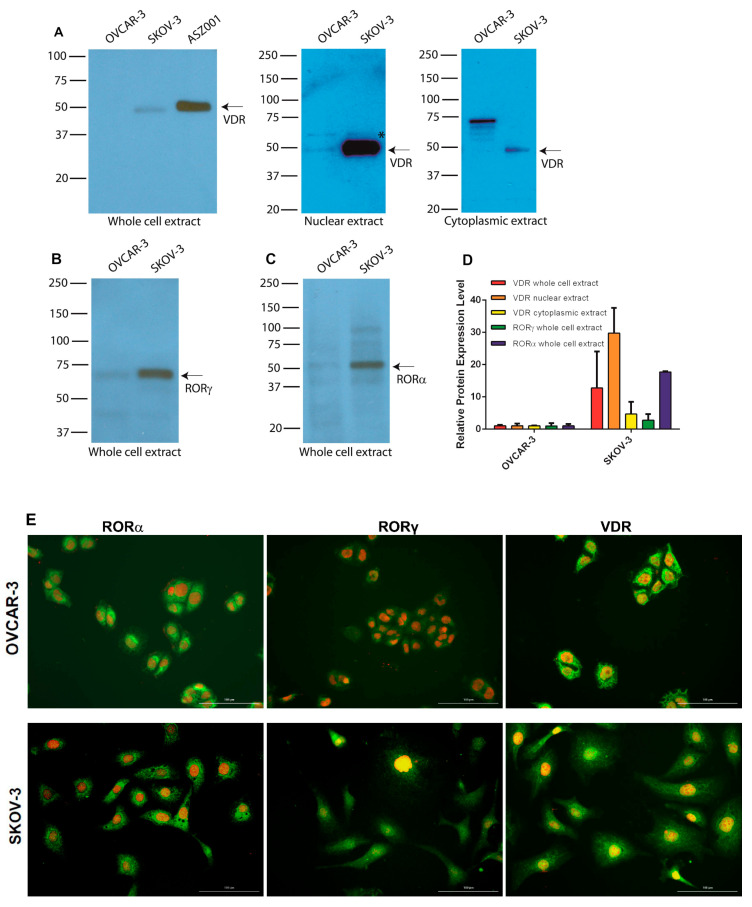
Figure 6.
RORα, RORγ, and VDR in OVCAR-3 and SKOV-3 ovarian cancer cells. Expression of VDR and RORα/γ in ovarian cancer cell lines as assessed by Western blot (WB): (A) VDR; (B) RORγ; (C) RORα. (D) Relative protein expression in OVCAR-3 and SKOV-3 as measured by WB in whole-cell extracts (n = 2) and nuclear (n = 2) and cytoplasmic (n = 2) fractions. ASZ001 (mouse basal carcinoma cell line) was used as a positive control for VDR expression in panel (A). (E) Immunofluorescence detection of RORα, RORγ, and VDR in OVCAR-3 and SKOV-3 ovarian cancer cells. Scale bars = 100 μm. * p < 0.05.

