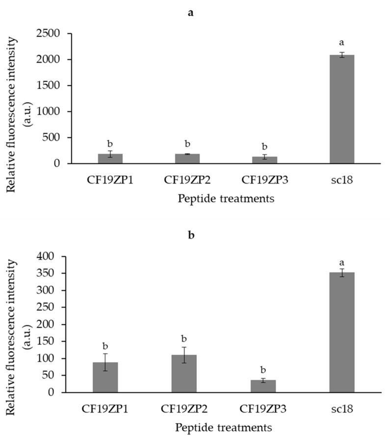
Figure 5.
Cellular uptake of peptides by HEK-293 (a) and HuH7 (b) cells as measured by flow cytometry. The cells were treated with 10 µM peptide for 1 h. The peptide sC18 was used as a positive control. Results are expressed as mean values ± standard deviation; n = 3. Superscript letters represent statistical differences among samples. Statistical differences were determined by Tukey’s test (p < 0.05).

