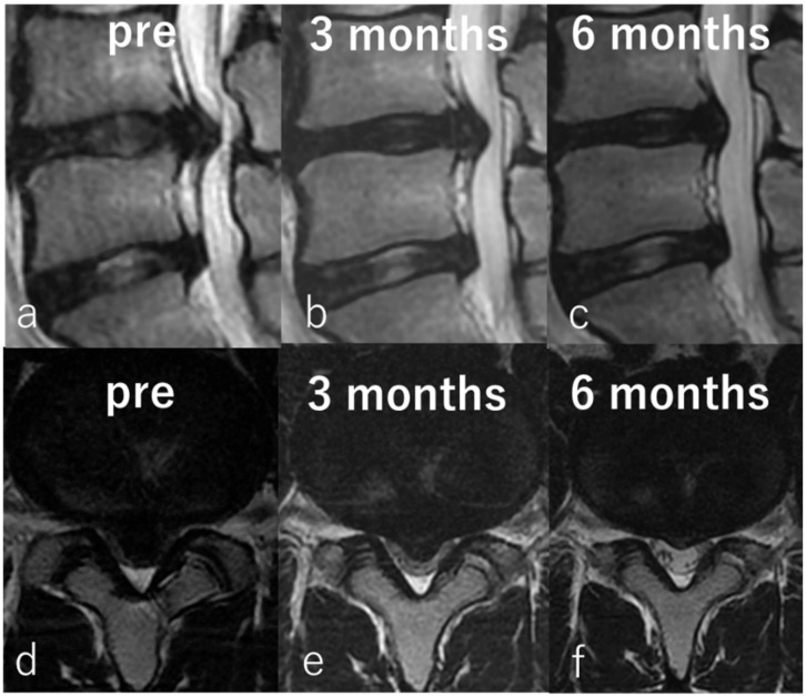
Figure 7.
Representative images of an 18-year-old man with large LDH. Preoperative sagittal (a) and axial (d) T2-weighted magnetic resonance imaging (MRI) showing sub-ligamentous LDH at L4/5. Sagittal (b) and axial (e) MRI taken 3 months after chemonucleolysis showing reduction of LDH. Sagittal (c) and axial (f) MRI taken 6 months after chemonucleolysis showing significant reduction in size.

