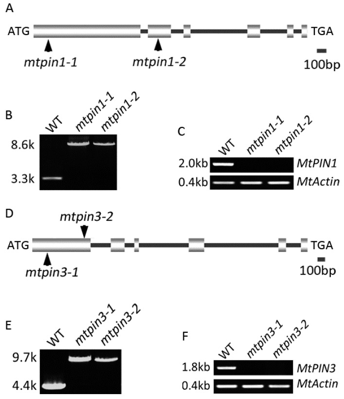
Figure 1.
Molecular characterization of MtPIN1 and MtPIN3 in M. truncatula. (A,D) Schematic representation of the gene structure of MtPIN1 and MtPIN3. The position of the ATG start and TGA stop codon are shown. Vertical arrows mark the Tnt1 insertion site in mtpin1 and mtpin3. (B,E) PCR amplification of MtPIN1 and MtPIN3 from wild-type (WT) and mtpin1, mtpin3 mutants. A single Tnt1 insertion (~5.3 kb) was detected in mtpin1-1, mtpin1-2, mtpin3-1, and mtpin3-2. (C,F) RT-PCR amplification of MtPIN1 and MtPIN3 transcripts in wild-type and mutants. MtPIN1 and MtPIN3 were not detected in mtpin1-1, mtpin1-2, mtpin3-1, and mtpin3-2. Actin was used as a loading control.

