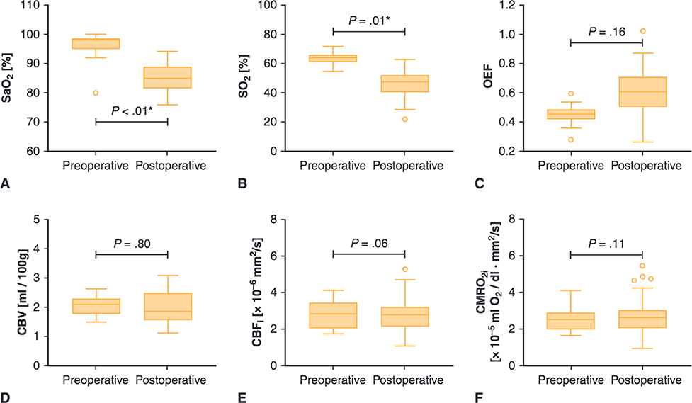
FIGURE 4.
Perioperative cerebral hemodynamics measures for patients with single-ventricle CHD. Upper and lower borders of boxes represent the first and third quartiles, middle line represents the median, upper and lower whiskers represent the maximum and minimum values within 1.5 * IQR (IQR is the interquartile range, or distance between the 1st and 3rd quartiles), and extra dots represent outliers. The statistical significance for comparisons is indicated with P values. *P values < .05. SaO2, Arterial saturation; SO2, cerebral oxygen saturation; OEF, cerebral oxygen extraction fraction; CBV, cerebral blood volume; CBFi, cerebral blood flow index; CMRO2i, cerebral oxygen metabolism index.

