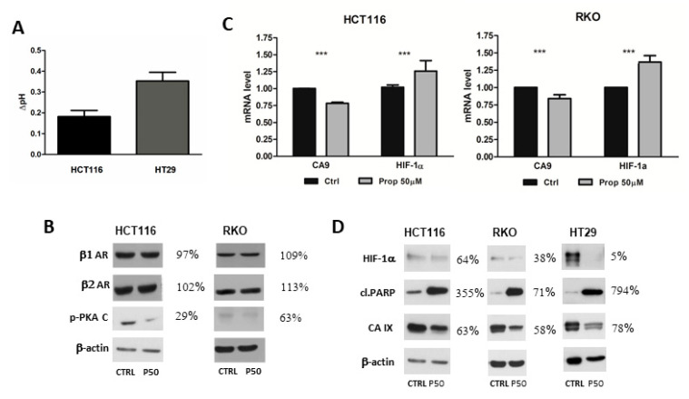
Figure 2.
Expression of β1 and β2-AR after beta-blockade by propranolol. (A) Propranolol mediated alkalization of extracellular pH in hypoxia. Colorectal cancer cells HCT116 and HT29 were treated with propranolol under hypoxic conditions for 24 h. Graph shows a difference between pH of propranolol treated samples and pH of appropriate controls (mean±standard deviation, n = 3) (B) Western blot analysis of β1 and β2-AR levels in HCT116 and RKO cells. No significant differences in the levels of β1 and β2 receptors were detected. Beta-blockade by propranolol decreased level of phosphorylated form of catalytic subunit of PKA under hypoxic conditions. (C) Graph of the results of quantitative PCR (qPCR) analyses. HCT116 and RKO cells were cultured in hypoxic conditions for 24 h. The expression of CA9 decreased after propranolol treatment. The mRNA level of HIF1α increased. The graph shows changes in mRNA levels normalized to actin. The results (mean ± stdev) represent the mean from three independent biological experiments, all performed in triplicates. Statistical significance was analyzed using the Student’s t-test and expressed as a p-value (*** p < 0.001) (D) Immunoblotting of CA IX and HIF1α proteins after treatment of colorectal cancer cell lines RKO, HCT116 and HT29 with propranolol under hypoxic conditions. Levels of CA IX and HIF1α in hypoxia decreased after beta-blockade in all three analysed cell lines and level of cleaved PARP increased. % values describe a difference in the signal of propranolol treated samples in comparison to the controls which were set as 100%. P50 represents propranolol at the concentration 50 µM.

