Figure 6.
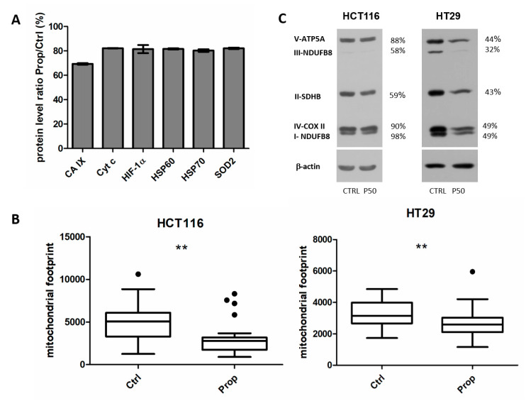
Propranolol reduces number of mitochondria and mitochondrial ox-phos enzymes. (A) Cell stress related proteins differentially expressed in spheroids after beta-blockade by propranolol. Using the Proteome Profiler Human Cell Stress Array Kit we analysed protein lysates from 15 spheroids from HCT116 cells treated for 5 days with propranolol and appropriate controls. The screening analysis showed 6 differentially expressed proteins. The results confirmed decreased levels of HIF1α and CA IX and of other proteins involved in mitochondrial metabolism and in oxidative phosphorylation. (B) Analysis of the amount of mitochondria using a Mito tracker MitoRed. Confocal analysis colorectal cancer cells monolayers cultured in hypoxia showed a decreased mitochondrial footprint, that is, the area of cell consumed by mitochondria signal, in samples treated by propranolol in both HCT116 and HT29 cells. Images were processed by MiNA toolset in ImageJ 1.41g. Box plot graphs of the measurements of at least 40 control and treated cells are displayed (** p < 0.01). (C) Western blot analysis of oxidative phosphorylation (OXPHOS) proteins in spheroid samples from HCT116 and HT29 cells. After propranolol treatment, the levels of proteins essential for the process of oxidative phosphorylation were reduced. % values describe a difference in the signal of propranolol treated samples in comparison to the controls set as 100%.

