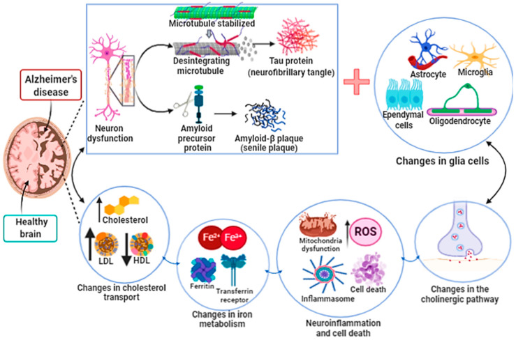
Figure 4.
Alzheimer’s disease. The development and progression of Alzheimer’s disease (AD) lead to atrophy, loss and dysfunction of both neurons and glial cells. AD begins in the dorsal raphe nucleus with subsequent progression to the cortex, which is the center of information processing and memory storage. The factors that promote the development of AD are still unknown. However, it seems that the intracellular accumulation in neurons of the phosphorylated Tau protein (neurofibrillary tangle) and the formation of amyloid-B plaque (senile plaque) in the extracellular environment and brain tissue both lead to neuron loss and dysfunction. In addition, the formation of neurofibrillary tangle and senile plaque alters the functions of glial cells, such as oligodendrocytes (responsible for the myelination of neurons), microglia cells (phagocytic cells) and astrocytes (responsible for the absorption and exchange of nutrients between neurons and blood vessels). Dysregulation of cholesterol transport and iron metabolism in the central nervous system contributes to poor prognosis of Alzheimer’s disease. All these associated factors lead to an increase in neuroinflammation and oxidative stress associated with mitochondrial dysfunction, compromising the production of ATP, altering the concentration of neurotransmitters in the synaptic cleft, finally promoting cell death.

