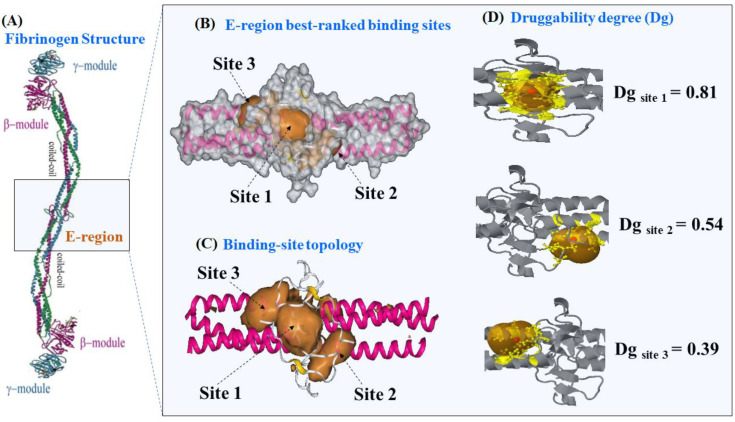
Figure 1.
(A) Details on the molecular structure of fibrinogen protein with the relevant regions, such as the two C-terminal portions of the carbohydrate-linking coiled-coil-like chains (γ-module and β-module) and the funnel hydrophobic cavity thrombin binding-domain (E-region, PDB ID: 1JY2 with 1.4 Å of resolution), the latter highlighted with the light-blue rectangle. (B) 3D-DCNN prediction of the three most relevant fibrinogen E-region binding-pockets (site 1, site 2, and site 3) depicted as van der Waals surfaces. (C) Representation of the binding-site topology linked to catalytic active sites, depicted as volumetric orange regions. (D) Prediction of the corresponding Dg for the best three catalytic binding sites of the fibrinogen E-region, depicted as orange transparent hull surfaces. Therein, the alpha sphere centers for predictions are shown as small red points within each site and surrounding target residues (yellow colored).

