Figure 6.
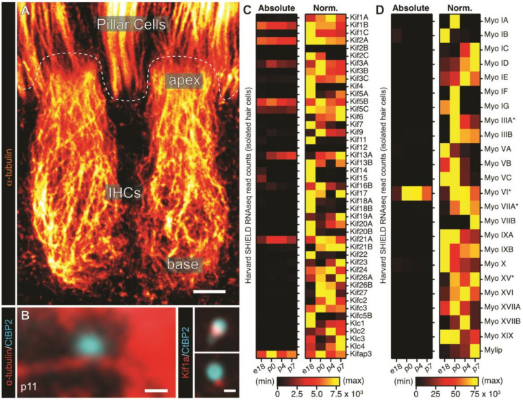
The role of cytoskeletal transport in IHC presynaptic AZ assembly. (A) The longitudinal microtubule network of murine inner hair cells (IHCs) is polarized in an apico-basal orientation. Shown is a super-resolution (STED) maximum projection of two p14 IHCs labeled against α-tubulin visualized with an intensity-coded look-up table, where warmer colors indicate higher intensities. (B) Representative STED images of cytoplasmically free-floating ribbon precursors (cyan) are located in close proximity to microtubule tracks (red) in IHCs prior to hearing onset (left panel) and colocalize with microtubule-based motor Kif1a (right panels). This suggests microtubule-based transport of ribbon material during development. (C,D) Developmental expression patterns of Kinesin motors (C) and Myosin motors (D), based on publicly-available RNA sequencing data of isolated murine IHCs replotted from the SHIELD database ([96]; https://shield.hms.harvard.edu/index.html). Illustrated are absolute expression patterns from embryonic day (e)18 to postnatal day (p)7 and the same data normalized to their maximum expression level over this time period to reveal temporal expression patterns of the individual targets. (*) indicates motor proteins linked with hereditary syndromic and/or non-syndromic hearing loss in humans (according to https://hereditaryhearingloss.org; October 2020). Scale bars: A 2.5 µm; B left panel: 250 µm, right panels: 200 µm. (B) with permission from Reference [4].

