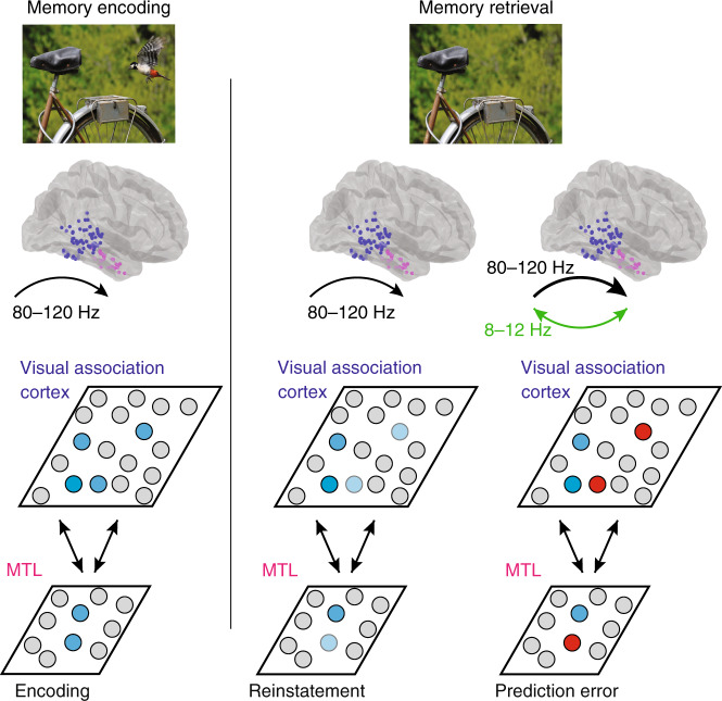
Fig. 7. Model of cortical–medial temporal lobe interactions during visuospatial recognition.
Initially viewing an image evokes image-specific 80–120 Hz activity within the visual association cortex and medial temporal lobe (MTL; blue units). During successful memory retrieval, the neural representation of a previously encoded image is reinstated within the visual association cortex and MTL. Successful reinstatement of this past experience is compared to current visual experience (dark and light blue units). In this case, the present visual experience no longer contains the bird that was in the remembered past visual experience. Any differences between present and past visual experience evoke an increase in 80–120 Hz power (bold arrow), which reflects an error signal, that arises in visual association cortex and propagates to the MTL (red units). This error signal is accompanied by low frequency coherence between these brain regions (green).

