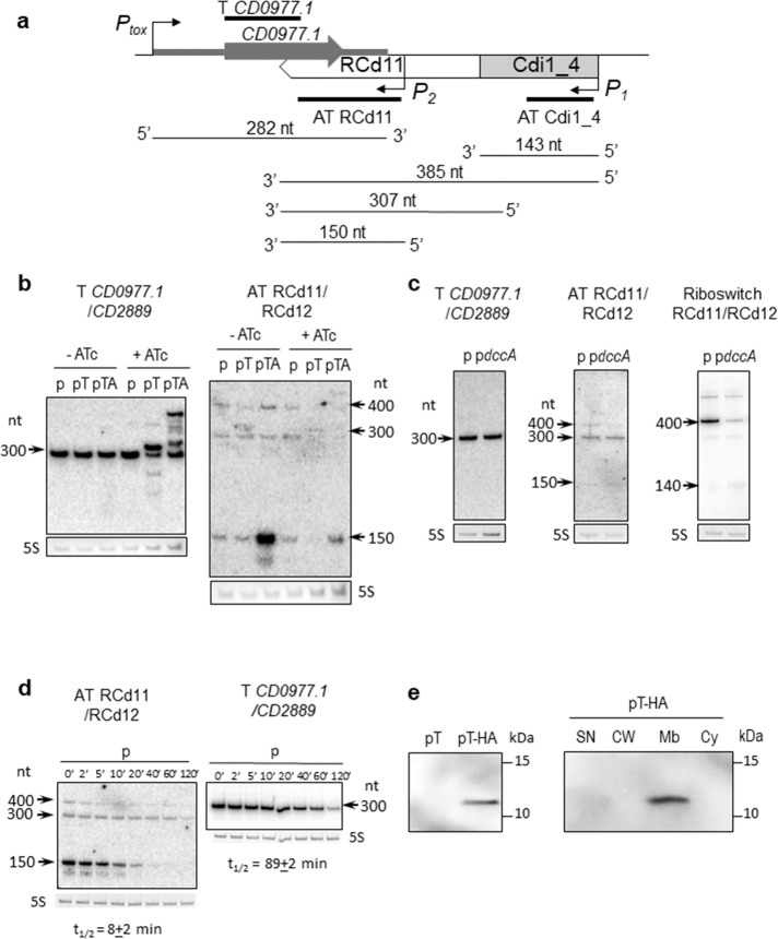
Fig. 2. Detection of CD0977.1 and RCd11 transcripts and CD0977.1-HA protein.
a A schematic of the CD0977.1-RCd11 TA pair genomic region and of the corresponding transcripts as identified by 5′/3′RACE and Northern blot. The Cdi1_4 riboswitch and the identified promoters are represented. The position of the different probes used in the Northern blot experiments is shown. b Northern blot of total RNA from C. difficile carrying p (empty vector), pT (expression of CD0977.1) or pTA (expression of CD0977.1 and its antitoxin) in the absence (−ATc) or in the presence (+ATc) of 250 ng/ml of the inducer ATc. c Northern blot of total RNA from C. difficile carrying p (empty vector) or pdccA (expression of the diguanylate cyclase encoding gene dccA) in the presence of 250 ng/ml ATc. d Northern blot of total RNA from C. difficile 630Δerm carrying an empty vector (wt/p) collected at the indicated time after addition of rifampicin. All Northern blots were probed with a radiolabelled oligonucleotide specific to the toxin (T CD0977.1/CD2889), the antitoxin (AT RCd11/RCd12) or the Cdi1_4/Cdi1_5 riboswitch (Riboswitch RCd11/RCd12) transcript and 5S RNA at the bottom serves as loading control. The arrows show the detected transcripts with their estimated size. The relative intensity of the bands was quantified using the ImageJ software. The half-lives for toxin and antitoxin transcripts were estimated from three independent experiments. e Detection and subcellular localization of the CD0977.1-HA protein. Immunoblotting with anti-HA detected a major polypeptide of ∼12 kDa in whole cell extracts of C. difficile carrying pT-HA (CD0977.1-HA) grown in the presence of 250 ng/ml of ATc but not in extracts of C. difficile carrying pT (non-tagged CD0977.1) (left panel). The culture of C. difficile carrying pT-HA was fractionated into SN supernatant, CW cell wall, Mb membrane, and Cy cytosolic compartments and immunoblotted with anti-HA antibodies. Proteins were separated on 12% Bis–Tris polyacrylamide gels in MES buffer. See Supplementary Figs. 6 and 7 for uncropped Northern-blots and Western-blots, respectively.

